
Figure 1: PRISMA Flow Diagram for Study Selection. The flowchart was designed according to the PRISMA guidelines 2020, showing study identification, screening, assessment eligibility, and final selection in the systematic review.
Irshad Khan1 | Asad Khalid2 | Mujtaba Ehsan2 | Muhammad Akram1 | Ejaz Ehsan Khan2* | Khaleeq Ullah3
1Department of Pathology, University of Lahore, Lahore, Pakistan | 2Department of Biosciences, COMSATS University, Islamabad, Pakistan
3Department of Pathology, University of Florence, Italy
*Correspondence: Ejaz Ehsan Khan (ahspreparations512@gmail.com)
Received: 15 January, 2026; Revised: 26 February, 2026; Accepted: 05 March, 2026; Published: 10 March, 2026
Background: Oxidative stress, mitochondrial dysfunction, and protein misfolding are some of the key and interrelated mechanisms in major neurodegenerative disorders. The purpose of this systematic review was to assess biomolecular therapeutic interventions of these pathways in the models of Alzheimer’s disease (AD), Parkinson’s disease (PD), and Huntington’s disease (HD). Methods: This is a systematic review that follow PRISMA 2020 guidelines. The searches took place up to from 2020 to 2026 in PubMed, Scopus, Web of Science, and Google Scholar. Experimental in vitro, in vivo, and ex vivo studies on biomolecular interventions that examine oxidative stress, mitochondrial dysfunction, or protein misfolding were accepted and reviews, editorials, conference abstracts, and non-English articles had been excluded. Screening and data extraction were done by two independent reviewers. The risk of bias was determined with the help of SYRCLE Risk of Bias tool of animal studies and Cochrane Risk of Bias 2.0 tool of in vitro studies and certainty of evidence was measured with the help of GRADE framework. Results: Twelve studies that passed the inclusion criteria were included following screening and eligibility. In disease models, interventions continually lowered the level of reactive oxygen species, restored mitochondrial bioenergetics, triggered Nrf2-mediated antioxidant response, lessened pathological protein aggregation, and neurobehavioral function. The risk of bias was low to moderate. Conclusions: The approach of the mitochondrial redox imbalance and proteostasis is a promising cross-disease treatment of neurodegeneration. To enhance the study further, future studies are needed to concentrate on standard experimental designs and clinical validation.
Keywords: Alzheimer Disease; Huntington Disease; Mitochondria; Neuroprotection; Oxidative Stress; Parkinson Disease; Protein Folding
Neurodegenerative diseases such as Alzheimer's disease, Parkinson's disease, and Huntington's disease were progressive and characterized by irreversible neuronal loss, progressive cognitive decline and progressive motor dysfunction. They represented a large public health concern around the world due to the demographic shift to age cohorts and a lack of curative therapies (e.g., synaptic dysfunction, protein aggregation, and energy metabolism failure) 1. A common pathological feature of these conditions was the disruption of mitochondrial integrity and redox imbalance by the excessive formation of reactive oxygen species (ROS) which compromises cellular integrity 2. Mitochondria were the energy-producing organelles of the cell and, consequently, the failure of this organelle leads to defective electron transport chain function, faulty autophagy-mediated removal (mitophagy), and altered calcium homeostasis, making neurons highly susceptible to oxidative injury and metabolic stress 3.
The main parameters that make up oxidative stress, mitochondrial dysfunction, and protein misfolding were interrelated processes that lead to the progression of the disease in neurodegenerative disorders 4. Oxidative stress occurred when a balance between oxidation and cell-based antioxidant mechanisms was upset by an increase in the formation of ROS, which led to lipid, DNA, and protein oxidation and the subsequent, direct chain injury to neuronal function 5. Mitochondrial dysfunction also enhances the production of ROS and impairs the cellular bioenergetic processes which lead to deficits of synaptic transmission and leads to neuronal death. These negative effects were consistently seen in many animal models of Alzheimer's disease, Parkinson's disease and Huntington's disease 6. Protein misfolding, such as of amyloid 2 and tau in Alzheimer's disease, of 2-synuclein in Parkinson's disease, and of huntingtin in Huntington's disease, imposed extreme stress on cellular quality-control processes. Such overloading not only saturates the ubiquitin-proteasome system and autophagic pathways, but also formed carefully driven cytotoxic protein filaments, which simultaneously enhance oxidative stress and exacerbate mitochondrial dysfunction 7.
Despite there had been extensive studies, there were still no effective disease-modifying therapies which were clinically effective. Several antioxidative and mitochondria-targeted strategies had shown poor translational results and this was largely due to poor mitochondrial engagement and insufficient mitochondrial integration into proteostasis pathways 8,9. Moreover, although proteinopathy-based interventions, including anti-amyloid-based ones, had partial ameliorative effects in the course of Alzheimer disease, they did not fully resolve the comorbid oxidative and energetic impairments, upon which disease progression depends 10,11. These limitations highlight the need of a systematic compilation of interventions that will e.g. deal with these biomolecular deficits in parallel in the central proteinopathies. This systematic review aims to critically discuss biomolecular treatments of oxidative stress, mitochondrial dysfunction and protein misfolding in Alzheimer's, Parkinson's, and Huntington's diseases. The review also intended to evaluate mechanistic pathways, therapeutic effects, possible risk of bias, and general certainty of the evidence, and establish future areas of translation and recommend future directions of disease-modifying strategies.
Figure 1: PRISMA Flow Diagram for Study Selection. The flowchart was designed according to the PRISMA guidelines 2020, showing study identification, screening, assessment eligibility, and final selection in the systematic review.
This systematic review followed the guidelines of the PRISMA 2020, as shown in Figure 1 12. A detailed systematic review protocol was developed, finalized, and verified internally prior to the commencement of data extraction to ensure research transparency and prevent reporting bias. A formal prospective registration code via external repositories was not sought for this specific cycle due to the rapid operational timeline.
Inclusion and Exclusion Criteria: Studies with defined research methods and data from adult NAFLD or patients with related liver characteristics were included. Studies including reviews, editorials or case reports in the evaluation were excluded.
Data Sources and Search String used: Eligible studies were original research investigations published as in vitro, in vivo or ex vivo studies reporting biomolecular therapeutic interventions that addressed oxidative stress, mitochondrial dysfunction or protein misfolding in the context of Alzheimer's disease, Parkinson or Huntington disease model. Studies were excluded if they were reviews, editorials, case reports, conference abstracts or non-English publications.
Study Selection and Data Extraction: Two reviewers (ME, MA) independently done screening of title, abstracts, and full texts and resolved any disagreements through discussion with a third reviewer (EEK). Data extraction was done using a standardized form for author, year, disease model, population characteristics, sample size, therapeutic intervention, molecular target/pathway, outcomes, and key findings. Two reviewers extracted data in separate sessions using a pre-developed data extraction form.
Primary Outcome and Quality Assessment: Risk of bias was determined by SYRCLE’ s Risk of Bias tool for animal studies and the Cochrane Risk of Bias 2.0 tool for in vitro studies 13,14. The level of certainty of the evidence was determined using the GRADE framework. The primary outcome was the effectiveness of biomolecular interventions in reducing oxidative stress, improving mitochondrial function, and mitigating protein misfolding across neurodegenerative disease models. Additionally, the study evaluated changes in key molecular markers (ROS levels, mitochondrial activity, and protein aggregation) to determine the overall therapeutic impact and mechanistic efficacy of the interventions.
The total number of records obtained as a result of the four database searches is 183 research articles: 55 records in PubMed, 36 records in Scopus, 25 records in Web of science, and 67 records in Google Scholar. After the removal of 98 duplicate articles, 85 different articles were left to screen titles and abstracts. After that, 38 were eliminated and at last 35 studies were eligible for assessment. Follow-up full-text evaluation led to the exclusion of 23 studies. Finally, 12 studies were included and met all inclusion criteria in the systematic review. Five of these were obtained in PubMed, one in Scopus, two in Web of Science, and four in Google Scholar. Table I is a summary of research that explored the Biomolecular Therapeutic Interventions on Oxidative Stress, Mitochondrial Dysfunction, and Protein Misfolding across Alzheimer’s, Parkinson’s, and Huntington’s Disease. Table I summarizes the study design, disease models, population characteristics, sample size, molecular target pathway, outcomes and key points in each study.
| Author (Year) | Country | Disease model | Study design/ Sample size/ population characteristics | Molecular target pathway | Outcomes | Therapeutic Intervention |
| Rodriguez et al., 2024 15 | USA | Alzheimer disease | Preclinical Experimental Study (In vivo study) n=12 mice, APP/PS1 (C57BL/6) mice in the experimental group, wild-type (C57BL/6) in control group | Mitochondrial oxidative stress and redox homeostasis, mitochondrial calcium uniporter pathway, and Aβ-mediated mitochondrial dysfunction pathway. | Improved mitochondrial redox ratio, reduced oxidative stress and Aβ burden, and modulation of mitochondrial calcium dysregulation. | SS31, Ru360, and Aβ immunodepleting. |
| Deng et al., 2024 16 | China | Alzheimer’s disease | Preclinical Experimental Study (In vivo) n=15 mice, experimental group (3×Tg-AD mice) and control group (C57BL/6 mice Wild-type) | KEAP1 inhibition disrupts the KEAP1–NRF2 complex | Artemisinin protected neurons from ferroptosis, reduced oxidative stress, and activated the KEAP1–NRF2–SLC7A11–GPX4 antioxidant pathway. | Artemisinin as a neuroprotective agent |
| Khodaei et al., 2020 17 | Iran | Alzheimer’s disease | Experimental, in vivo. n= 60 rats total Male Sprague Dawley rats. | Mitochondrial dysfunction, oxidative stress, NF-κB signaling, ROS production, sirtuin family activity. | Decreased oxidative stress markers (ROS), restored cortical ATP levels, enhanced cytochrome c oxidase activity. | Ellagic acid |
| Chen et al., 2025 18 | China | Alzheimer’s disease | Preclinical experimental intervention (in vitro) n=18 Double transgenic APP/PS1 mice, C57BL/6J wild‑type mice | Aβ aggregation Microglial polarization Apoptosis pathway Mitochondrial function Energy metabolism | Reduced Aβ accumulation, Shift microglia to anti-inflammatory phenotype, Apoptosis decrease, increase Mitochondrial membrane potential and ATP production and neuroprotection | Photo biomodulation (PBM) |
| Eo et al., 2024 19 | South Korea | Parkinson’s disease | Preclinical Experimental Study (In Vivo + In Vitro) For in vitro n=64 mice C57BL/6 mouse. For in vivo neuronal cell models | Mitochondrial dysfunction, oxidative stress, neuroinflammation, and dopaminergic neuron loss. | Motor behavior, dopaminergic neuron survival, microglial activation, and pro-inflammatory cytokines improve Parkinson's. | Mitochondrial transplantation (PN‑101 isolated from human mesenchymal stem cells) |
| Kim et al., 2022 20 | South Korea | Parkinson’s disease | Preclinical Experimental Study (In Vivo + In Vitro) For in vitro n=60 mice B6; C3-Tg, B6C3F1/slc mouse For in vivo SH-SY5Y | α‑synuclein aggregation, mitochondrial dysfunction, proteostasis, and oxidative stress pathway. | Motor behavior, dopaminergic neuron loss, α-syn aggregates, microglia activation, transcriptome changes | MT101‑5 Neuroprotective mitochondrial therapy |
| Liu M et al., 2023 21 | India | Parkinson's disease | In vitro experimental study Human neuroblastoma SH-SY5Y cells | ROS modulation, Mitochondrial membrane potential maintenance, Apoptosis pathway, Neuron protection, and Dopaminergic neuron marker. | Prdx-2 protects SH-SY5Y cells from MPP⁺ toxicity by reducing oxidative stress and stabilizing mitochondria. | Prdx-2 provides neuroprotective therapy. |
| El‑Shamarka et al., 2023 22 | Egypt | Parkinson’s disease | In vivo experimental study n=70 mice Swiss albino mice exposed | Molecular targets: oxidative stress, dopaminergic neurons, and glial activation | Oxidative stress was reduced, glial activation decreased, and motor behavior improved. | Curcumin, Levodopa /Carbidopa, Rasagiline, Combination therapies |
| Jiang et al., 2025 23 | China | Huntington’s disease | Combined in vivo (mouse) and in vitro (PC12 cells) experimental study n=100 mice, C57BL/6J mice | Nrf2/HO-1 pathway, Oxidative stress (ROS), Synaptic proteins, | Oxidative and neuronal damage, NaHS protected via Nrf2 activation | NaHS (H₂S donor), ML385 (NRf2 inhibitor) |
| Sun X et al., 2020 24 | China | Huntington's disease | Preclinical in vitro experimental study n=60 mice R6/2 transgenic mice, Wild-type littermate mice. | Reduction in oxidative stress, inhibits mutant huntingtin aggregation. | Reduction in mutant huntingtin aggregates and polyQ inclusions lowered ROS. | Ellagic acid |
| Gao et al., 2021 25 | China | Huntington’s disease | Experimental in vivo study HEK293, IMR90 Human cell lines | ROS pathways, mitochondrial bioenergetics. | Plant extracts targeted oxidative stress, protein aggregation, and protein quality control pathways | Ethanolic extracts, Acetone extracts, Quercetin |
| Cordeiro et al., 202126 | USA | Huntington’s disease | In vitro experimental study 50 worms per group Transgenic C. elegans strains (Bristol N2, AM141, AM101, HA759, CL2070, CF1553) | ROS pathways, mitochondrial function, and neuronal metabolism. | Oxidative stress biomarkers, mitochondrial activity, and neurobehavioral measures. | Antioxidant/nutritional therapy. |
| ROS = Reactive oxygen species, NF-κB = Nuclear factor kappa-light-chain-enhancer of activated B cell, ATP = Adenosine triphosphate ,MMP = Mitochondrial membrane potential, ELISA = Enzyme-linked immunosorbent assay, Nrf2 = Nuclear factor erythroid 2–related factor 2, HO-1 = Heme oxygenase-1, Keap1 = Kelch-like ECH-associated protein 1, SLC7A11 = Solute carrier family 7 member 11, GPX4 = Glutathione peroxidase 4, Aβ = Amyloid-beta, Cytc = Cytochrome c, LC3II = Microtubule-associated protein 1A/1B-light chain 3-II, PKM2 = Pyruvate kinase M2, PGC1 = Peroxisome proliferator-activated receptor gamma coactivator 1-alpha, CD86 = Cluster of differentiation 86, ARG1 = Arginase 1, SH-SY5Y = Human neuroblastoma cell line, HE = Haematoxylin and eosin staining, NaHS = Sodium hydrosulfide (H₂S donor), 3BP = 3-bromopyruvate | ||||||
Pharmacological and genetic treatment in transgenic and toxin-induced murine and rodent models of Alzheimer disease had the effect of reducing mitochondrial reactive oxygen species. They reestablished cellular redox balance, triggered Nrf2-dependent antioxidant signaling and ameliorated amyloid -associated mitochondrial dysplasia, leading to an increase in ATP production and reduced ferroptotic cell death. In models of Parkinson disease, therapeutic regimens reduced oxidative stress, preserved mitochondrial membrane potential, deferred the formation of alpha -synuclein aggregates, reduced neuroinflammatory responses, and produced a higher motor performance. In the studies the therapeutic agents reduced the reactive oxygen species, magnified Nrf2/HO signaling pathways, reduced mutant Huntington aggregates, optimized mitochondrial bioenergetics, and enhanced autophagy-mediated clearance of pathogenic proteins all in the disease models of Huntington.
In all the twelve studies where design-specific tools such as SYRCLE RoB for animal studies and Cochrane Risk of Bias 2.0 tool for in vitro studies were used, the overall risk of bias was low to moderate, as shown in Figure 2. Two animal model-based studies showed a high risk of bias due to lack of blinding of outcome assessment and incomplete outcome data. However, none of the studies were assessed as having critical risk of bias across all domains. Consequently, the overall GRADE certainty of the evidence was moderate.
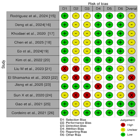
Figure 2: Traffic plots for evaluating risk of bias assessment in vitro studies using Robvis tool.
Overall, the included studies demonstrated consistent therapeutic effects across Alzheimer’s, Parkinson’s, and Huntington’s disease models, particularly in reducing oxidative stress and improving mitochondrial and proteostatic function. Most studies showed favorable molecular and functional outcomes, although methodological limitations contributed to low-to-moderate risk of bias. The moderate certainty of evidence supports the potential of targeting these interconnected pathways while highlighting the need for further standardized and clinically relevant investigations.
The Neuroprotective interventions based on biomolecular therapeutic intervention approaches to oxidative stress, mitochondrial dysfunction, and protein misfolding show a consistent outcome in every experimental model of Alzheimer’s disease, Parkinson’s disease, and Huntington disease. In models of Alzheimer’s disease, interventions led to a decrease in the generation of reactive oxygen species by mitochondria, a reestablishment of redox homeostasis, an increase in antioxidant signaling via Nrf2, an increase in ATP production, and a decrease in the amyloid-associated mitochondrial impairment 27,28. Convergence of mechanisms was also similar in Parkinson’s disease models, where therapies maintained mitochondrial membrane potential, decreased α -synuclein aggregation, inhibited microglial activation, and enhanced motor responses especially through mitochondrial-targeted agents and mitochondrial transplantation approaches 29,30. Activation of the Nrf2/HO -axis, decrease of mutant huntingtin aggregation, mitochondrial bioenergetic recovery, and improvement of autophagic clearance was repeatedly reported in Huntington disease models 31.
These findings were consistent with growing levels of translational evidence that oxidative stress and maladaptive mitochondrial quality control increase protein aggregation and neuronal susceptibility in AD, PD and HD 32,33. The recent experimental and preliminary translational research highlighted the Nrf2 activation, mitophagy amplification, and ferroptosis regulation as the potential cross-disease treatment mechanisms 34. These mechanistic pathways were supported by our results especially the key role of Nrf2-mediated antioxidant defence and mitochondrial stabilization 35,36. Moreover, the behavioral improvement that was observed in transgenic models is in line with the preclinical meta-analyses indicating that mitochondrial redox system-targeting can cause eventual functional recovery 37. Notably, the consistency of the results between diverse models supported the claim that multi-target biomolecular treatments can have a wider disease-modifying capability than single-pathway models 38. Nonetheless, in spite of the mechanistic consistency, the variability in the dosing schedules, period of intervention and outcome measures restricts the direct comparability and the necessity of standardized preclinical frameworks 39.
The limitations need to be considered as various databases did not target grey literature and unpublished data in a systematic manner, which could have contributed to the bias of publication and the registration of the protocol was not conducted and this can be constraining transparency and reproducibility. Also, experimental models and result reporting were heterogeneous, and thus not able to undergo quantitative synthesis, requiring a narrative approach. Studies were mostly preclinical in vitro or animal studies and as such lack the direct clinical translatability. The sample sizes were often small and reporting on randomization, allocation concealment, and blinding procedures was not always clear, which would pose higher chances of performance and detection bias. Cross-study comparison was further limited by variability in the models of diseases, the timing of the interventions and biomarkers. Furthermore, the long-term safety and pharmacokinetics were not adequately covered in a number of experiments, as well as dose-response relationships.
Possible improvements in future research would include standardized experimental design, transparent reporting, in accordance with ARRIVE guidelines, and multisite preclinical validation in order to enhance reproducibility 40. It can be expected that a combination of multi-omics profiling and mitochondrial functional assessments will help understand the mechanistic pathways and finding predictive biomarkers. Translational research that would provide the gap between preclinical and early-phase clinical research is needed, especially on Nrf2 activators, mitophagy modulators and ferroptosis-targeting agents. Lastly, a more disease-modifying potential in neurodegenerative diseases could be provided by simultaneous mitochondrial dynamics, proteostasis, and oxidative stress combinational therapeutic approaches.
This systematic review highlights the overlapping role of the oxidative stress, mitochondrial dysfunction, and protein misfolding in Alzheimer disease, Parkinson disease, and Huntington disease as well as critically appraises biomolecular therapeutic solutions to these interrelated conditions. In preclinical in vitro and in vivo models, the interventions always decreased reactive oxygen species, recovered mitochondrial bioenergetics, triggered Nrf2-mediated antioxidant systems, enhanced the proteosome and exhibited a neuroprotective and behavioural advantage. On the whole, it is possible to assert that mitochondrial-redox modulation is a promising cross-disease treatment approach, but it needs a thorough translational research and uniform preclinical models to enhance reproducibility and bring these interventions to the clinical stage.

AL-NAJAM Journal of Medical and Life Sciences, Al Najam J Med Life Sci 2026:1(1), p6-11 (anjmls.org) © 2026 Authors. This work is published by Multidisciplinary Scholarly Advancement and Research MSAR Institute. The full terms of Journal Publishing policy is available at https://anjmls.com/index.php/anjmls/publishing-license and incorporate the Creative Commons Attribution – Non Commercial (CC BY, NC 4.0) License https://creativecommons.org/licenses/by-nc/4.0/. By accessing the work you hereby accept the Terms. Non-commercial uses of the work are permitted without any further permission, provided the work is properly attributed. Publisher’s Note: MSAR Institute remains neutral with regard to jurisdictional claims in published maps and institutional affiliations, and assumes no liability for the scientific accuracy or clinical efficacy of the content herein, as they rest entirely with the authors.