Hamayun Saqib1*
1Department of Clinical Medicine, Jiujiang University of Jiangxi, China
*Correspondence: Hamayun Saqib (hamsaqibch@gmail.com)
Received: 01 May, 2026; Revised: 01 June, 2026; Accepted: 05 June, 2026; Published: 10 June, 2026
Alzheimer’s disease (AD) is the most common cause of dementia in the world, and it is marked by deterioration of cognitive functions; the formation of amyloid-β in the brain; and tau, the formation of tau pathology; synaptic degeneration; and local susceptibility, especially in the hippocampus and association cortices. The most common processes identified to cause neuronal injury and disease development are oxidative stress and mitochondrial dysfunction. Ultraweak photon emission (UPE), a spontaneous biophotonic emission in response to oxidative metabolic reactions, has been linked to possible cellular redox imbalance and neural activity. Since oxidative changes in AD are region-specific, UPE can indicate region-specific neurodegeneration. Nevertheless, existing research on brain UPE in AD is insufficient and primarily focuses on global oxidative indicators, rather than regional patterns. Lack of systematic analysis of the region-specific UPE changes is also a major gap in research. This aims to examine the regional differences in brain UPE in Alzheimer’s, and determine whether they correlate with oxidative imbalance and neuropathological severity. The detection of specific UPE signatures could contribute to the enhanced comprehension of spatial disease heterogeneity and aid in its advancement as a possible functional biomarker of the early disease detection and progression tracking.
Keywords: Alzheimer's Disease, Dementia, Oxidative Stress, Neurodegenerative Diseases, Mitochondria
Alzheimer's disease (AD) is a progressive neurodegenerative condition that is characterized by cognitive, memory, and functional deterioration, mostly in the geriatric population. AD is the most prevalent type of dementia that affects people globally, with great social and economic costs 1. Extracellular amyloid-beta (Aβ) plaques and intracellular neurofibrillary tangles made of hyperphosphorylated tau protein are the neuropathological hallmarks of AD, and they all lead to the impairment of synapses, neuronal loss, and brain network activity. Although much work has been done, there are still unclear data on the exact mechanisms of the regional vulnerability in AD, which explains the necessity to develop new strategies to identify and define the initial pathological alterations. According to recent findings, ultra weak photon emission (UPE) or biophoton emission can be used to detect a sensitive index of oxidative stress and mitochondrial dysfunction in the brain, which is core to AD pathophysiology 2.
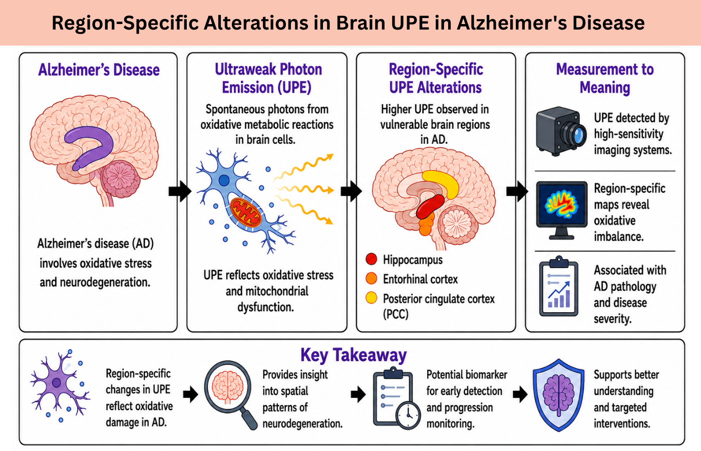
UPE is the spontaneous release of low-intensity photons by living cells during metabolic processes, especially as by-products of reactions of reactive oxygen species (ROS). Since neuronal tissues have high metabolic capacity and are prone to oxidative stress, their UPE can be measured and used as a non-invasive measure of biochemical changes on the cellular scale. Hence, the analysis of the UPE variations in the brain regions provides a promising avenue to get early biomarkers and to have insight into the spatial heterogeneity of neuro-degeneration in AD. The brain is not homogeneously impacted in AD, with some areas being more susceptible than others; i.e., the hippocampus, entorhinal cortex, and the posterior cingulate cortex are vulnerable, while others are relatively resilient 3.
Atrophy of the hippocampus, such as that, is linked to memory loss, but cortical involvement is linked to more general cognitive impairments. The mechanisms of this regional specificity are one of the basic questions of AD studies. The role of oxidative stress as one of the major mediators of neuronal vulnerability has become accepted. Overproduction of ROS, dysfunction of the mitochondrion, lipid peroxidation, and oxidation of proteins all decrease the integrity of the neurons and the functions of the synapses 4. Since UPE is strongly related to ROS-mediated biochemical activities, the specific changes in UPE in different regions could be an indication of the disparate oxidative load within the different regions of the brain in AD. The findings of the experiments confirm the possibility of UPE being a biomarker of neurodegenerative diseases. The in vitro studies have shown that neurons exposed to oxidative stress levels are characterized by high levels of UPE, which is linked to mitochondrial dysfunction and apoptotic signaling 5.
Regional differences in UPE in animal models of AD have also been shown, with the hippocampal and cortical neurons having higher rates of emission compared to other regions that are less affected by disease-related amyloid and tau pathology. These findings suggest that UPE cannot only be a prognostic variable but can also be employed to monitor disease progression and response to treatment therapy 6. Besides basic oxidative stress, the amyloid pathology and the mitochondrial dysfunction may act in the mechanism of region-specific UPE changes. It is also known that Aβ aggregates impair mitochondrial respiratory chains, favor the development of ROS, and disrupt calcium homeostasis, which subsequently leads to local energy deficiency and neuronal injury. Similarly, tau pathology also contributes to oxidative stress and synaptic dysfunction in specific groups of neurons, with regional variability 7. The cumulative product of these pathological processes may, therefore, be measured with UPE measurements and provide insight into the original biochemical environment of the AD-involved brain areas.
The latest technology has allowed a more accurate identification of UPE on biological tissues and with greater sensitivity. The quantitative mapping of UPE in live brain tissue and cultured neurons can now be performed by photomultiplier tubes, charge-coupled devices, and new imaging systems. These techniques allow comparative studies across brain regions and experimental models, which help show the spatially different patterns of oxidative stress 8. High-resolution mapping such as this is of particular importance in AD, where early detection of even subtle regional dysfunction has the potential to enhance the accuracy of prognostic analysis and direct therapeutic interventions. Besides, UPE research in AD can have a wider implication on the dynamics of the disease over time. Longitudinal studies indicate that oxidative stress and UPE changes are antecedents of overt neurodegeneration and cognitive impairment, which is in line with the oxidative stress hypothesis of AD. Early detection of UPE changes in the region would thus allow preclinical intervention to be set in place, which may slow down or prevent further loss of neurons. As well, a combination of UPE information with standard neuroimaging technologies, including MRI and PET, can present a multimodal solution to identifying the interaction between structural degeneration, amyloid/tau load, and oxidative metabolic dysfunction 9.
The difficulties in the translation of UPE research into clinical applications persist even in spite of encouraging evidence. The variability of the intensity of emissions, technical shortcomings of the methods to detect the ultra-weak signals in the living organism, and the impact of confounding factors (age, sex, metabolic condition, etc.) should be taken into consideration 10. To achieve reliability and reproducibility, standardization of measurement protocols, calibration of detection systems, and rigorous testing in human cohorts are necessary. However, there is a promising future for UPE as a non-invasive biomarker of region-specific neurodegeneration, which is novel and promising in AD research.
In brief, Alzheimer's disease has a strong regional susceptibility, and there is selective involvement of hippocampal, cortical, and limbic structures. The focus of this selective neurodegeneration is oxidative stress and mitochondrial dysfunction, and ultraweak photon emission is a sensitive measure of these biochemical disruptions. It has been observed that UPE changes in the brain in a region-specific manner are indicators of the spatial heterogeneity of oxidative damage and are associated with characteristic AD pathology. The development of UPE detection technology has provided the opportunity to develop early biomarkers, disease longitudinal follow-ups, and to integrate with other traditional neuroimaging. Further investigation of UPE in AD is likely to improve our knowledge of disease pathogenesis, enable early diagnosis, and give targeted therapeutic interventions to maintain neuronal functioning and mental abilities.
Conclusion: The regional changes of ultraweak photon emission (UPE) indicate that the brain regions involved in Alzheimer's disease are not equally affected by oxidative stress and mitochondrial dysfunction. Recent studies indicate that UPE could be a useful functional biomarker for the early detection of neurodegenerative changes and the progression of disease. The development of new UPE detection technologies has improved the potential for integration with traditional neuroimaging techniques. UPE should be validated in clinical trials to confirm its reliability as an early diagnostic, prognostic, and targeted therapeutic monitoring tool in Alzheimer's disease.

AL-NAJAM Journal of Medical and Life Sciences, Al Najam J Med Life Sci 2026:1(2), p4-6 (anjmls.org) © 2026 Authors. This work is published by Multidisciplinary Scholarly Advancement and Research MSAR Institute. The full terms of Journal Publishing policy is available at https://anjmls.com/index.php/anjmls/publishing-license and incorporate the Creative Commons Attribution – Non Commercial (CC BY, NC 4.0) License https://creativecommons.org/licenses/by-nc/4.0/. By accessing the work you hereby accept the Terms. Non-commercial uses of the work are permitted without any further permission, provided the work is properly attributed. Publisher’s Note: MSAR Institute remains neutral with regard to jurisdictional claims in published maps and institutional affiliations, and assumes no liability for the scientific accuracy or clinical efficacy of the content herein, as they rest entirely with the authors.